Testis, also known as testicle, in animals, the organ that formssperm, the male reproductive cell, and androgens, the male hormones. In humans the testes occur as a Couple of oval-shaped organs. They’re contained inside the pocket sac, that is found directly behind the erectile organ and before of the anus.
Anatomy of the Testes
In humans both testis weighs around 25 grams and are 4–5 cm long and 2–3 cm in diameter. Each is covered by a fibrous capsule called the tunica albuginea and is divided by partitions of fibrous tissue from the tunica albuginea into 200 to 400 wedge-shaped sections, or lobes. Within each lobe are 3 to 10 coiled tubules, called seminiferous tubules, which produce the sperm cells.
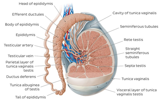
The partitions between the lobes and therefore the body fluid tubules each converge in one space close to the anal facet of every testis to make what’s known as the cavum testis.The testes contain germ cells that differentiate into mature spermatozoa, supporting cells known as Sertoli cells, and testosterone-producing cells known as Leydig (interstitial) cells. The germ cells migrate to the vertebrate testes from the embryonic food sac. The Sertoli cells that are interspersed between the germinal animal tissue cells inside the body fluid tubules are analogous to the granulosa cells within the ovary, and therefore the Leydig cells which are located beneath the tunica albuginea, in the septal walls, and between the tubules, are analogous to the hormone-secreting interstitial cells of the ovary. The Leydig cells are irregularly shaped and commonly have more than one nucleus. Frequently they contain fat droplets, pigment granules, and crystalline structures; the Leydig cells vary greatly in number and appearance among the various animal species. They are surrounded by numerous blood and lymphatic vessels, as well as by nerve fibers.
Ovary Anatomy
The ovaries are female reproductive organs that are related to the testes in men. They produce the ova (eggs), which develops into a fetus when it becomes fertilized, they also create the female sex hormones estrogen and progesterone. There are two ovaries, both are located within the pelvic area beside the uterus (womb).
Ovarian Structure
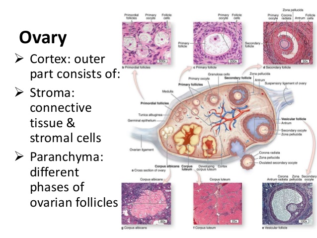
The ovaries are oval-shaped and are round about 1 1/2 inches long. They are pinkish-gray in color and have an uneven surface. The ovaries are connected to the uterus by the fallopian tubes, or oviducts, which carry the eggs into the uterine cavity. Each ovary contains varied Graafian follicles, egg-containing tubes that grow and develop between pubescence, sexual maturation, and climacteric, once the monthly cycle stops. Once agirl is fertile, every month a follicle travels to the surface of the ovary, bursts, associated releases an egg and its fluid contents into a Fallopian tube.
The Graafian follicles are mounted during a network of supporting tissue (stroma) and blood vessels.They are covered by a clear, smooth, plasma-like membrane that develops from the peritoneum—lining of the abdominal cavity. Additionally within the ovaries are tiny numbers of corpus lutea—the remains of Graafian follicles that have discharged an egg and are within the method of being reabsorbed by sex gland tissue. Every month the endocrine gland (the connective tissue of a Graafian follicle) is chargeable for the assembly of progestogen. Progesterone is the pregnancy hormone that readies the lining of the uterus for the arrival of a fertilized egg.