- Ribosomes are ribo-nucleo-protein particles found in almost all cells.
- These are the assembly shops for protein synthesis. So they are described as protein factories.
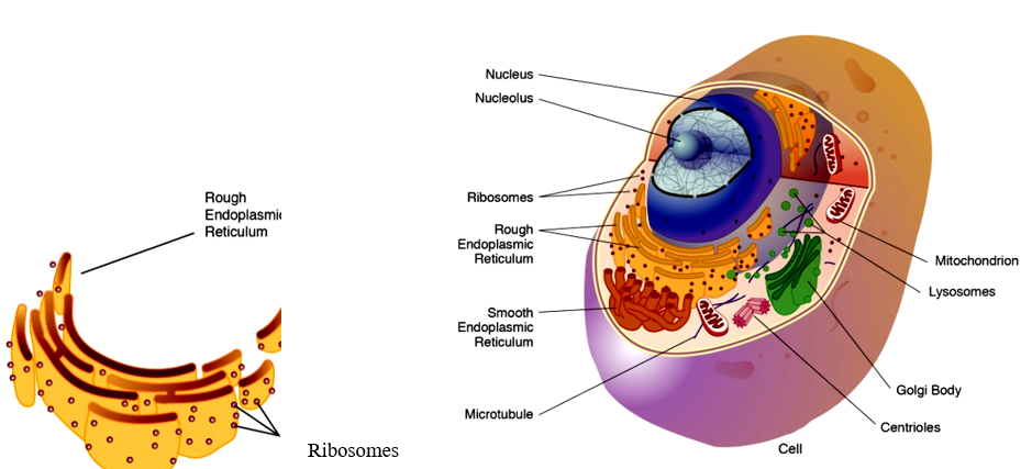
- They are found in the cytoplasm or attached to the endoplasmic reticulum.
- Ribosomes were first observed by Claude and named them as microsomes.
- George Palade (1953) named them as ribosomes.
Ribosomes
Structure & Composition
- Ribosomes are spherical bodies composed of ribonucleic acid (RNA) and proteins and are not surrounded by any membrane.
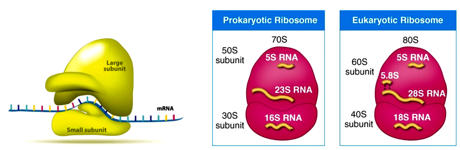
- Each ribosome has two subunits, namely a large subunit and a small subunit.
- In eukaryotes, the ribosome is of 80S type consisting of 60S and 40S subunits.
- The prokaryotic ribosomes are 70S (made of 50S and 30S subunits).
- Here ‘S’ (Svedberg’s Unit) stands for the sedimentation coefficient; it is indirectly a measure of density and size.
- The subunits occur separately in the cytoplasm.
- They join together to form ribosomes during protein synthesis.
- Generally 5 or more ribosomes line up and join in an mRNA chain.
- Such a small subunit mRNA chain string of ribosomes are called Polyribosome or Polysome.
Functions
- Protein synthesis.
- The mRNA passing through the ribosome is protected from nucleases.
- Ribosomes protect newly synthesised polypeptide chains from proteases enzymes.
Cytoskeletal structures
It is a system of protein filaments in the cytoplasm of all eukaryotic cells. It gives the cell shape and the capacity for directed movement. Cytoskeletal structures consist of small tubules and filaments.
Three types of cytoskeletal filaments have been recognised,
- Microtubules
- Microfilaments and
- Intermediate filaments.
Each type of filament is formed of different protein subunits.

Microtubules
- These are unbranched, hollow, elongated cylindrical organelles found in the cytoplasm. They are about 25 nm in diameter.
- They are made up of spirally arranged globular subunits of proteins called tubulin.
- In nerve cells microtubules give rigidity.
- Spindle fibres formed during cell division are the aggregation of microtubules. After breaking up of the tubules, they have the capacity to reassemble at any part of the cell.

Functions
- Intracellular transport.
- It determines the shape of the cells.
- Sliding movements of the spindle microtubules help in movement of chromosomes during cell division.
- It helps in cell mobility
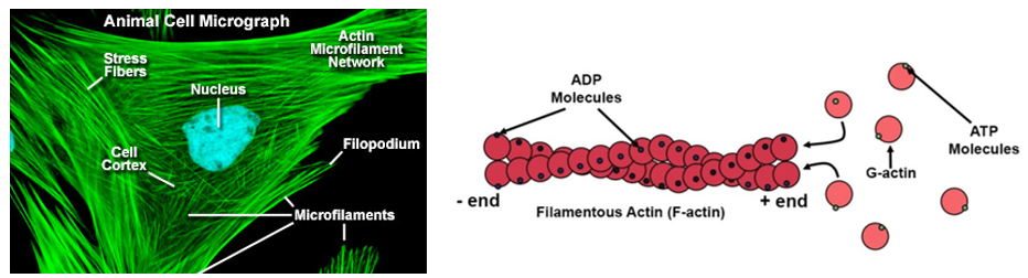
Microfilaments
These are very fine protein filaments that are 5 to 7nm in diameter and occur in bundles.
- They are composed of subunits called actin.
- These are the most abundant cytoskeletal elements in many eukaryotic cells.
- It forms the core of microvilli and a crucial component of the muscle cells.
Functions
- They help in the movement of cell organelles.
- They are involved in cytoplasmic streaming movements.
- They are responsible for muscle contraction.
- They are involved in endocytosis and exocytosis.
Intermediate filaments
These are filaments of intermediate size ranging from 8 – 10 nm in diameter (intermediate between microtubules and microfilaments). They are made of globular proteins such as Keratin, Vimetin, Desmin etc.
Cília and Flagella
The cilia (cili – eye lash) and flagella (little whip) are microscopic, contractile, filamentous processes of the cytoplasm which move the entire cell or organism, act as sensory organs and perform many mechanical functions of the cell.
- Cilia and flagella are structurally almost identical.
- Both are composed of 11 longitudinal fibres (9 + 2).
- 2 are running through the centre, while the
- Other 9 are around the centrally placed ones

- Each fibril is composed of two subfibril units.
- Each central fibril is singlet fibre and each outer fibril is doublelet fibre.
- A basal body is seen below each cilium or flagellum in the cytoplasm.
- The portion that projects from the free surface of cell is called shaft or cilium.
Functions of cilia and flagella
- Locomotion
- Cilia help in capturing food in lower aquatic animals.
- Ciliary movements help in the elimination of the solid particles from respiratory tract
- They act as sensory organs.
Difference between Cilia and Flagella
- Flagella are longer than cilia.
- Cilia have no restriction in number. But flagella occur singly or a few in number
- Flagella occur at one end of the cell, while the cilia may occur throughout the surface of the cell.
- The flagella beat independently, while the cilia tend to beat in a co-ordinated rhythm
- The flagella exhibit undulatory motion, while the cilia move in a sweeping or pendular stroke.