The male and female gametophytes differ in a way such that the pollen grain or microspore produces the male gametophyte while the megaspore produces the female one. These two are the stages of heterosporous plants.
The male gametophyte.
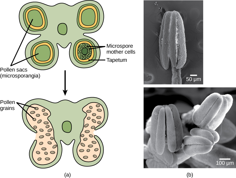
In an immature anther, the male gametophyte develops. The male reproductive system of a plant contains a structure called microsporangium where the development of pollen grains takes place. The microspores develop into pollen grains in the microsporangia. They are found in the anther located at the stamen’s end.
Meiosis occurs in the microsporangium where the microspore mother cell divides to form four microspore where all of them are used to form a pollen grain. These microspores are nourished by an inner wall called tapetum hence contributing key components to the pollen wall. Upon the maturity of the pollen grains two cells called the generative cell which is contained in the larger pollen tube cell and the pollen tube cell. The tube cell forms the pollen tube where the generative cell migrates to enter the ovary upon germination. Two male gametes during the division of the generative cell inside the pollen tube. Hence the pollen are released from the anther when the microsporangia bursts upon maturity.
Female Gametophyte.
The development of the female gametophyte vary between species but the overall development contains two phases.
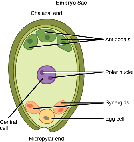
The first process called megasporogenesis is where the single cell in the diploid megasporangium goes through meiosis to form four megaspores where only one survives. The second phase is called megagametogenesis where the surviving haploid megaspore goes through mitosis to form an eight-nucleate, seven- cell female gametophyte called megagametophyte or embryo sac. The polar nuclei move to the equator, fusing to form a single diploid central cell, which later fuses with a sperm forming a triploid endosperm. This leads to the formation of the antipodal cells when the nuclei position themselves on the end of the embryo sac opposite the micropyle leading to their degeneration. The nucleus which is the closest to the micropyle becomes the female gamete (egg cell) and the other two adjacent become the synergid cells. The synergid cells function to guide the pollen tube for fertilization hence disintegrate afterwards. After successful fertilization,the fertilized ovule forms the other tissues of the seed and the diploid develops into the embryo.
The megasporangium is protected by a double layered integument which later protects the embryo sac. This double layered integument develops into the seed coat after fertilization to protect the seed while the ovule wall becomes part of the fruit. The integument does not close completely leaving an opening called micropyle where the pollen tube enters the female gametophyte for fertilization.