Dicotyledonous Leaf (Dorsi-ventral leaf)
A thin cross section of dicot leaf shows three main parts, namely epidermis, mesophyll and vascular system.
Epidermis
- Epidermis covers the entire surface of a leaf.
- It is covered by a thick layer of cuticle.
- It is single layered and composed of closely packed cells.
- In some plants like Ficus, the epidermis is multilayered.
- In Xerophytes, the outer walls of epidermal cells are thickened to prevent water loss.
- In water plants, the leaves possess a waxy covering to prevent wetting of leaves.
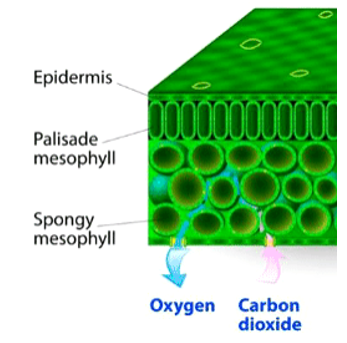
- The abaxial (lower) surface of leaf bears more stomata than the adaxial (upper) surface.
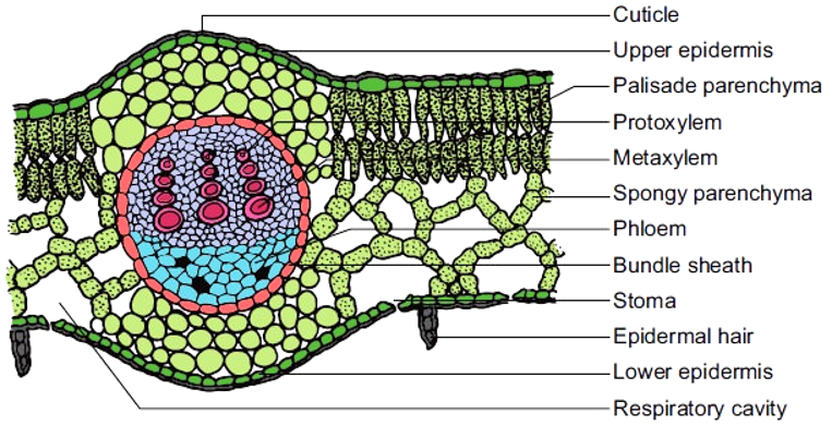
Mesophyll
- Tissue between the upper and lower surface is called mesophyll.
- It forms the ground tissue of a leaf and is composed of parenchyma cells.
- The upper side mesophyll is composed of single layer of narrow elongated cells arranged at right angles to the epidermis. It consists of large amount of chloroplasts. It is called the palisade layer.
- The lower part is made of several layers of isodiametric parenchyma cells, with large intercellular spaces. It is called the Spongy layer.
Vascular bundles
- The vascular tissue in leaves occur in the form of veins and the midrib.
- Size of the bundles depends on the size of the veins.
- The bundle is surrounded by a compact layer of parenchyma cells called the bundle sheath.
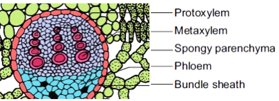
- The vascular bundle is collateral closed.
- The xylem is seen at the upper side, and phloem is found in the lower side.
- Protoxylem is seen towards the upper side, and hence it is pseudo-exarch condition.
- Phloem consists of sieve tubes, companion cells and parenchyma cells.