1. Fertilization
Fertilization is the union of the female gamete (egg) and the male gamete (spermatozoa). Whether it occurs naturally inside the female reproductive system or with the assistance of reproductive technologies outside of the human body, the product is a structure called a zygote. When a woman is ovulating she releases one egg into her Fallopian tubes.
Following spermatozoa ejaculation inside the vagina, special secretions help them to swim through the cervix towards the uterine tube where fertilization takes place within 24-72h.The fertilized egg or zygote then begins to move toward the uterus, as the cells divide into the next stage, a blastocyst.
2. Blastocyst Development
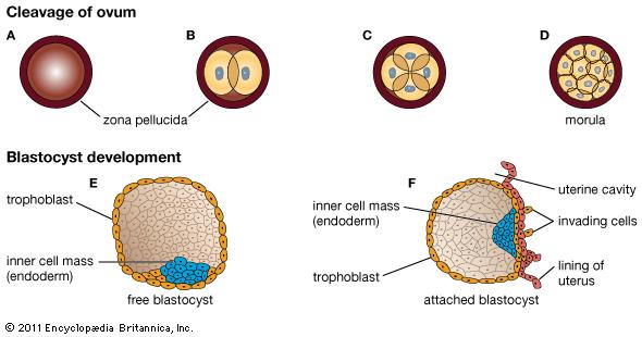
Soon after fertilization, the embryo is created from a small group of cells that are constantly dividing inside of a complex structure called the blastocyst. It is formed by two groups of cells, inner and outer cells, and fluids. The blastocyst stays inside a protective cover during maturation called zona pellucida, which could be described as an egg shell. The outer cells are located right below this cover, which will create the future placenta and surrounding tissues to support fetal development in the uterus. The inner cells of the blastocyst will become the different tissues and organs of the human body, such as bones, muscles, skin, liver, and heart.
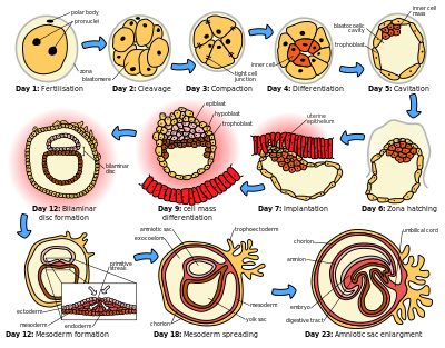
The cells within the blastocyst grow fast, they go through many changes and convert into more specialized cells, making the structure very tight. In humans, these changes happen during the first few days of development, before the implantation in the uterus. At this stage blastocyst is released and broke by the zona pellucida. It moves through the Fallopian tubes towards the uterus and implants around day ten.
3. Blastocyst Implantation
When the blastocyst reaches the uterus it implants in the endometrium, the mucus membrane which lines the uterus. The external cells of the blastocyst and the uterine inner lining, together, will create the future placenta. The placenta is a system through which nutrients are transferred to the baby to removes his/her wastes.
4. Embryo Development
As the blastocyst reaches the final steps in the implantation process into the inner lining of the uterus, it evolves into a structure called an embryo. This is the time when internal organs and external structures develop. The mouth, lower jaw, throat are emerging, while the blood circulation system starts its evolution and a heart tube is created. The ears arise and arms, legs, fingers, toes, and eyes are being shaped. Brain and the spinal cord are already made up, but the digestive system and sensory organs begin their flourishment. The first bones are replacing the cartilage.
Placenta formation
This hollow ball of cells moving through the uterus is the blastocyst, searching for an implantation site. Its outer layer beginning to extend out and implant in the uterine lining, searching for the uterine blood vessels that would nourish it throughout the pregnancy. As it went deeper, a single layer of cells from the mother’s uterine lining surrounded it, so that it would be protected from harm. On Day 9, as it grew larger and more complex, the blastocyst became an embryo. Here it’s about the size of a pinhead. Also on Day 9, the outer layer of the embryo developed spaces called lacunae. The lacunae filled up with blood from the mother’s uterine lining. On Day 13, small projections from the embryo’s chorionic layer reached out into the uterine lining. The chorionic layer is one of the membranes that surround the embryo and help it implant. On Days 15 through 21, blood vessels began to form beneath this chorionic layer. Around Day 21, the embryo’s blood stream and the mother’s blood stream were in such close contact that nutrients and oxygen could cross from mother to embryo. This was how the embryo first got its food and air from the mother, and technically this is when the placenta began to function and formed.